
Combining endoscopy and ultrasound to detect what standard tests may miss — for earlier diagnosis and better care
If you are searching for the best Endoscopic Ultrasonography (EUS) in Max Hospital Dwarka, Dr. Lovkesh Anand offers expert diagnostic care using advanced EUS technology. This minimally invasive procedure provides highly detailed images of the digestive tract, pancreas, bile ducts, and surrounding organs, helping in early and accurate diagnosis of complex gastrointestinal conditions.
Endoscopic Ultrasonography (EUS) is a specialized procedure that combines endoscopy and ultrasound imaging to examine internal organs with exceptional clarity. At Max Hospital Dwarka, Dr. Lovkesh Anand uses EUS to evaluate conditions affecting the esophagus, stomach, pancreas, liver, bile ducts, and lymph nodes.
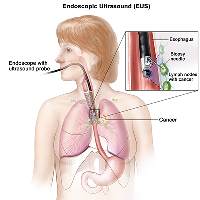
A thin, flexible tube with an ultrasound probe at its tip is gently inserted through the mouth or rectum, depending on the area being examined. This probe generates high-frequency sound waves to produce real-time, high-resolution images from inside the body, often more detailed than those from CT or MRI scans
Insertion of the tube – A thin, flexible endoscope with a miniature ultrasound probe at its tip is used for the procedure.
Route of insertion – Depending on the area being examined, the scope is carefully inserted either through the mouth (for stomach, pancreas, bile duct, or esophagus) or the rectum (for rectum and lower digestive tract).
Ultrasound technology – The probe emits high-frequency sound waves, which bounce off internal organs and tissues.
Real-time imaging – These sound waves are converted into detailed, high-resolution images on a monitor, allowing doctors to visualize organs and structures that cannot be seen clearly with regular endoscopy.
Greater accuracy – EUS provides much sharper and closer views compared to CT or MRI scans, especially for small tumors, cysts, or blockages.
Guided procedures – If needed, doctors can also perform a fine-needle biopsy during EUS to collect tissue samples without surgery.
Pancreatic tumors or cysts – EUS can detect even very small growths or cysts in the pancreas that might be missed on CT or MRI. It also helps doctors decide whether the cyst is benign (non-cancerous) or cancerous, guiding the right treatment.
Bile duct stones or blockages – Gallstones or narrowing in the bile ducts can block the flow of bile, causing severe pain, jaundice, or infection. EUS provides high-resolution images to confirm the blockage and plan further treatment like ERCP.
Esophageal, gastric, and rectal cancers – EUS plays an important role in cancer staging. It shows how deeply the cancer has spread into the wall of the digestive tract and whether nearby lymph nodes are involved, helping doctors plan surgery or therapy.
Unexplained abdominal pain – When regular ultrasound, CT, or endoscopy cannot explain persistent abdominal pain, EUS allows a closer look at internal organs like the pancreas, bile ducts, and stomach wall to detect hidden causes.
Chronic pancreatitis – In patients with repeated inflammation of the pancreas, EUS shows scarring, calcification, or narrowing of ducts. This helps in assessing disease severity and planning long-term management.
Enlarged lymph nodes or masses – Swollen lymph nodes or abnormal lumps inside the chest or abdomen can be examined with EUS. In many cases, a fine-needle biopsy can also be done during the same procedure to check for cancer or infection.
Minimally invasive with no external incisions – EUS is performed using a thin, flexible scope, so there are no cuts or scars. This makes the procedure safe, less painful, and recovery much quicker compared to traditional surgery.
Highly accurate diagnosis of tumors and lesions – EUS provides very clear, close-up images of internal organs, helping doctors detect even small tumors, cysts, or growths that may not be visible on CT or MRI.
Can guide fine-needle aspiration (FNA) for biopsy or fluid sampling – During the procedure, doctors can insert a tiny needle through the scope to collect tissue or fluid samples. This allows accurate diagnosis of cancer, infection, or other conditions without open surgery.
Reduces the need for exploratory surgery – Since EUS offers both imaging and biopsy in one procedure, patients often avoid major exploratory surgeries, lowering risks, costs, and recovery time.